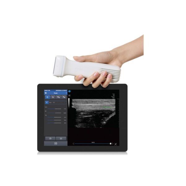
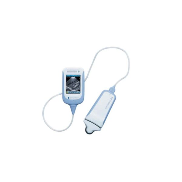
The POCKETPRO H2 500L linear wireless handheld ultrasound system is designed for MSK, vascular access, and procedural guidance.
Range of 6 to 12 MHz
Color, PW, M-mode functions
Customizable presets
JPEG and DICOM file formats
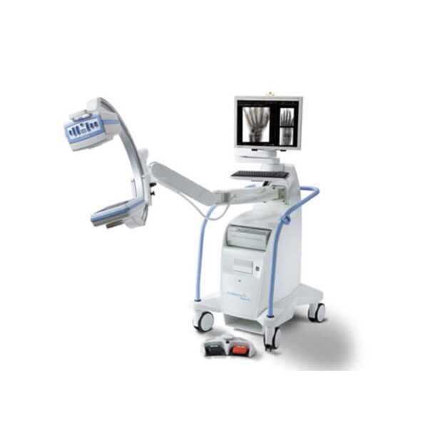
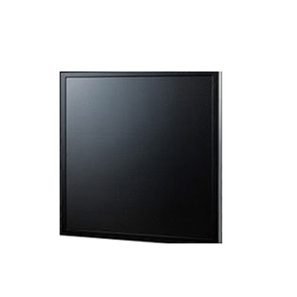
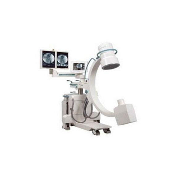
Hologic continues to raise the bar for high quality fluoroscopy imaging with our CsI (cesium iodide) CMOS flat detector and its exclusive rotating capabilities. The CsI material provides high sensitivity, resulting in lower dose and high quality images.
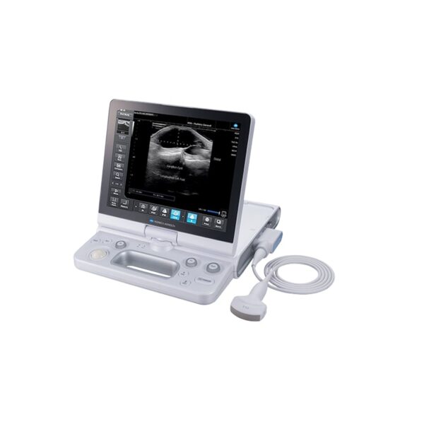
The SONIMAGE HS1, with its high sensitivity and broadband linear probe, features several advancements in transducer technology, such as a multi-level matching layer, optimized component materials, and nanofabrication technology. SONIMAGE HS1 uses a five-level wave control algorithm to generate separate harmonic signals that cover the entire receive spectrum of the system. As a result, SONIMAGE HS1 overcomes many common trade-offs between resolution and penetration, providing the right balance for optimum, best-in-class image quality.
The SONIMAGE P3 is a true portable ultrasound machine that gives clinicians the ability to do more for patients where and when they need it most – at the point-of-care. With its small footprint and weighing less than a pound, this handheld device can accelerate and improve interventions and decision making time.
Intuitively designed like a smart-phone, the P3 equips users with a non-invasive tool to see what’s beneath the surface in patients, giving care providers’ quicker access to information and improving patient care. The SONIMAGE P3, cost-effectively empowering physicians to do more with less – providing them imaging solutions in real time.
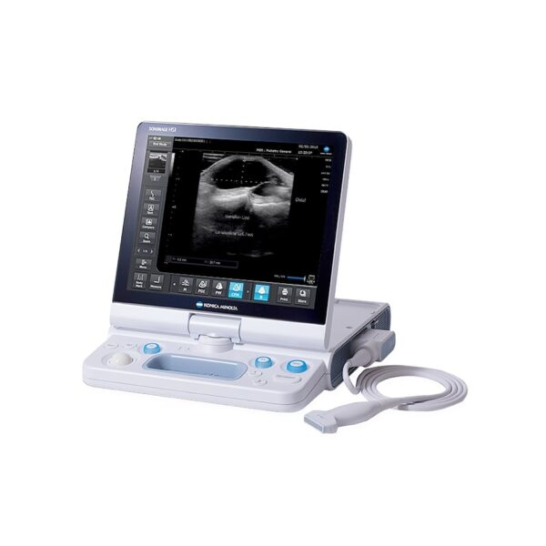
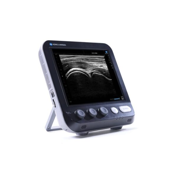
Introducing the next-generation, compact point-of-care ultrasound system, the SONIMAGE® MX1 Platinum Ultrasound System, with detail resolution for superior image quality.
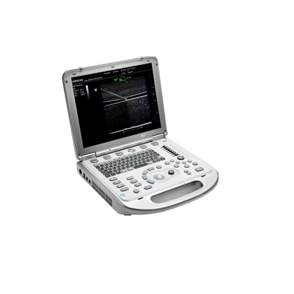
The M7 Diagnostic Ultrasound System is designed to meet the needs of clinicians’ busy and challenging point-of-care environments. With M7’s crystal clarity and crisp, clear image quality, it can perform any exam, from abdominal to vascular to cardiac, with efficiency and accuracy. Just by choosing a transducer, the M7 brings you more benefits in more ways than ever with wellness within reach.
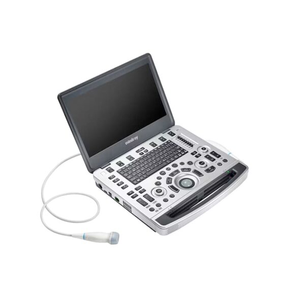
Based on Mindray’s new generation ultrasound platform, mQuadro, M9 has raised the industry standards to an all new level. Advanced signal transmission and reception processors provide highly sensitive and accurate echo detection. Innovative transducer technologies allow for better penetration, higher resolution, greatly enhancing your diagnostic experience.
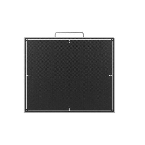
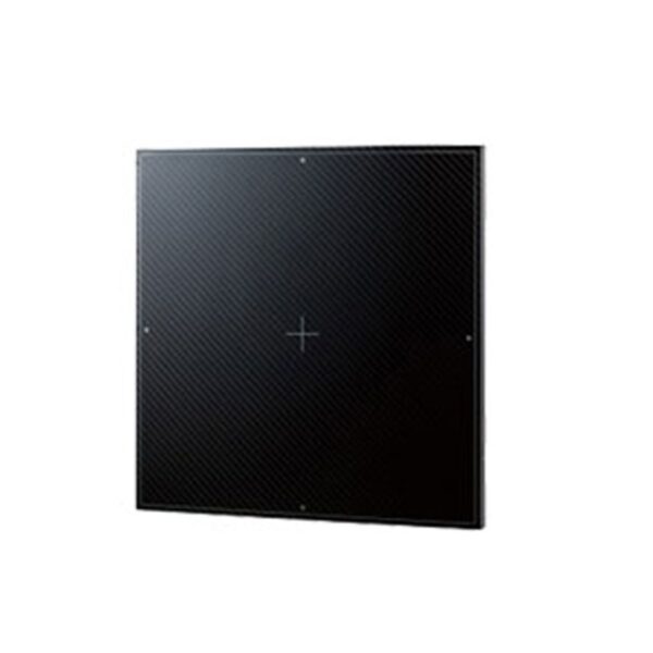
Tiger DR-Cesium X-Ray Panels are cassette size flat panel detectors. TigerView DR-Cesium X-Ray Panels fit into existing bucky trays. The entire active area is prepared with the “Autosense” technology and takes care of the exposure timing. This ultra thin and lightweight wireless detectors allow digitizing without modification of existing conventional imaging systems. Cable free wireless option gives the user a true wireless detector at a fraction of the cost.
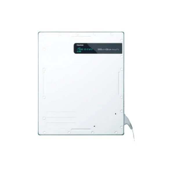
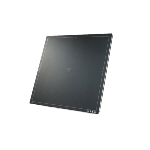
The FDR D-EVO has achieved 384 ×460mm size and 14mm thick which is equivalent to a CR cassette. The other main characteristics of the FDR D-EVO are 2.8kg 1weight, minimum 3seconds preview time and minimum 9seconds cycle time. Since the size is equivalent to a CR cassette, it is possible to load the FDR D-EVO into an existing upright/table X-ray system and can be handled in the same fashion as a CR cassette.
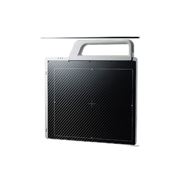
This lightweight, wireless digital detector was created through the technological know-how of Rayence, the global leading manufacturing of digital imaging detectors. The uniqueness of this versatile detector solution includes an ergonomically designed, removable handle that can be attached to either the portrait or landscape location of the detector frame for ease of use.
For added flexibility, the 1012WCA can also be used as a tethered detector. With this configuration, the battery will recharge without it having to be removed and placed into a separate charger.
Experience the convenience, versatility and flexibility available only in the 1012WCA.
Automatic Exposure Detection
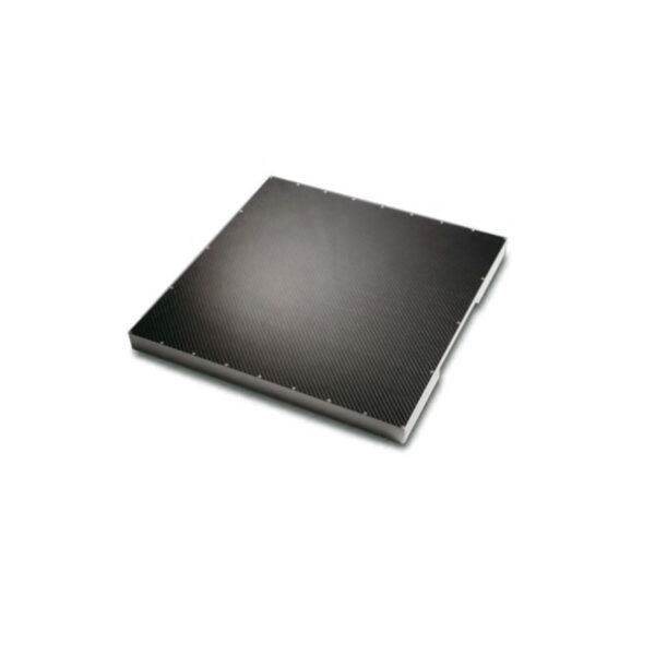
Single 17 x 17 inch a-Si TFT PIN Diode
Fixed Flat Panel Detector
Control Box with 30 ft. Power Cord
9 or 21ft. Panel Cable
NOTE: Detectors DO NOT include Grid or Cabinet
Housings (Optional 200 Line Grids are recommended
for Human use with Alto DR Detectors.)
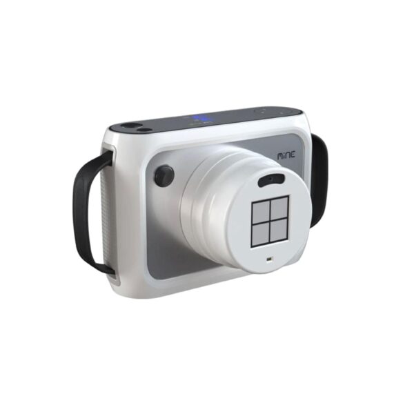
Associated X-Ray Imaging offers you a full line of radiology x-ray imaging systems that provide state-of-the-art performance to meet your patient care needs. The OTOM Mine Alnu is an FDA approved* handheld, wireless x-ray generator which delivers 80kv weighing less than 6 pounds. This device is ideal for orthopedics, sports medicine, podiatry, and veterinary.
– Single Function Foot Switch
– Real Time Processing
– Filters/Windowing/Rotation/Mirroring
– Post Processing-Edge Enhancement
– Rotation, Windowing, Inverse, Image Crop
– High Frequency X-ray Generator 20,000 Hz
– Iris and Slot During live Fluoroscopy
– Fluoroscopic Operation from 40kV to 110kV at 0.2-Maximum 6ma and Pulse Modes
– Radiographic Operation from 40kV to 110kV at Maximum 20ma
– 8ma Mode for Digital Radiograph/Snap Shot Mode
– Dual Mode 9/6 in. Image Intensifier
– High Contrast Camera
– Body Region and Application Specific Key, Extremities, Chest, head, Spine, Hip Metal, Soft Tissue, 1/2 Dose
– Large Patient Diameter key
– Integrated Cable Pusher Allow for Easy Maneuverability and Positioning
– Two 18 in. High Resolution/Brightness TFT Flat Panel Monitors, 1280×1024
– 1280x1024Vx12 bit Highline Video Image Display with Dual Graphic Overlays
– 360 Degree Digital Continuous Image Rotation
– Last Image Hold
– Invert, White on Black
– True 1024 Shades of Gray
– Real Time Noise Reduction (Low, Medium, High)
– Snapshot, Electronic Noise Reduction by Multiple Frame Integration Preset for Better Image Quality
– Touch Screen Text Keyboard for Patient Annotation
– Auto Store Feature
– True 16 bit Image Processing and Storage
– 10,000 Digital Image Stored in 1Kx16 bit Image Display
The Exa® Platform provides the infrastructure for managing data across the imaging workflow
Integrated RIS/PACS/Billing platform
Diagnostic Zero Footprint (ZFP) viewer with Server-Side Rendering
Real-time performance dashboard for maximum efficiency
How we do it.
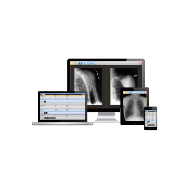
Efferent’s platform of cloud-based solutions transforms the speed and efficiency of healthcare delivery with streamline processes, rapid communication, and robust resources. 100% Cloud-based imaging platform – NO Servers, No Software, Use Any Device. Imagine connecting and automating healthcare giving you more….
TigerView offers workflow specific to your market. Defining market needs and developing a PACS product affordable for the consumer is always the underlying focus for a Televere Systems product.
Advanced Diagnostic Tools – Manipulate crystal clear images
Easily review angles and measurements, find the mid-line and center point, utilize stitching, cropping and magnification tools which help to better diagnose and communicate with your patients.
Market Specific Measurements – More Efficient workflow
Multitude of measurement tools specific to all markets such as Medical, Podiatry, Chiropractic, Veterinary, Non-Destructive Testing and Security.
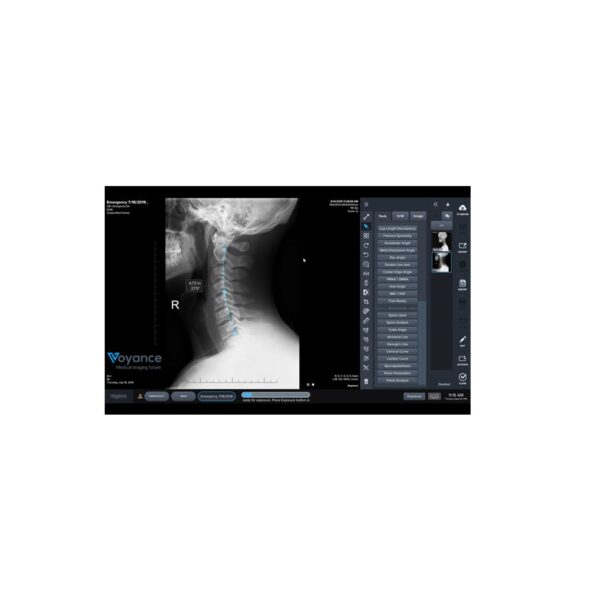
Voyance VPACS Server is an affordable solution for viewing, sharing and storing DICOM images for the long term. Purchased as a stand-alone solution on dedicated hardware, or as part of an Acquire/Archive DR solution, VPACS provides practices with a scalable, upgradable solution to archive x-ray image data and comply with HIPAA guidelines. VPACS server securely shares patient data throughout your local network and allows providers to review images in any room.
Chiropractic clinics seek to present a professional and efficient image to their clients. The Opal-CHIRO PACS solution for these clinics allows the patients’ x-rays to be quickly imported into the system, available for diagnosis much quicker than film. Thus the system also affords the clinic faster reads, reduced or eliminated film costs, and eliminates the need for physical storage space to archive film.